Author(s):
In 2025, the National Institutes of Health (NIH) announced that the agency would no longer provide funding for research grants that only include animal models in their proposed studies.1 The need for alternative preclinical testing has proved necessary, as close to 90% of drugs that pass animal testing do not end up being approved by the FDA due to safety and efficacy issues in humans.2 Importantly, based on a 2020 study in the Journal of the American Medical Association, the total cost to bring a drug to market, considering the rate of drug failures, is $1.3 to $2.8 billion,3 and the time required to proceed from preclinical studies to the bedside can range from 10 to 15 years or more.
The Cawley Center for Translational Cancer Research (Cawley Center) at the Helen F. Graham Cancer Center and Research Institute has established a solid tumor organoid core to generate patient-derived human tumor models for preclinical drug screening and translational research. Recent studies show that mouse models do not adequately reflect human cancer genetics and immune responses, contributing to poor translation of preclinical findings. In contrast, patient-derived organoids provide a cheaper, faster, and more biologically relevant model system. Organoids can also provide an advantage over animal models since they can be used to study interactions on a single-cell level in real-time and provide the ability for high-throughput analyses of tumors from patients with diverse genetic backgrounds.4 Therefore, the use of patient-derived tumor models makes it possible to design personalized treatment strategies in the future, offering more durable responses to therapy.
What Are Organoids?
Organoids, or mini tumors, mimic body tissues by providing artificial extracellular matrices and three-dimensional (3D) architecture for patient-derived tumors; they can allow for the culture of multiple cell types in a more natural environment than traditional cell culture. Unlike traditional 2D culture models, organoids preserve the histopathologic and genetic characteristics of human tumors. Organoid cultures can be supplemented with growth factors and additional cell types to model key components and interactions within the tumor. In addition, organoids are relatively simple to maintain and expand in culture.
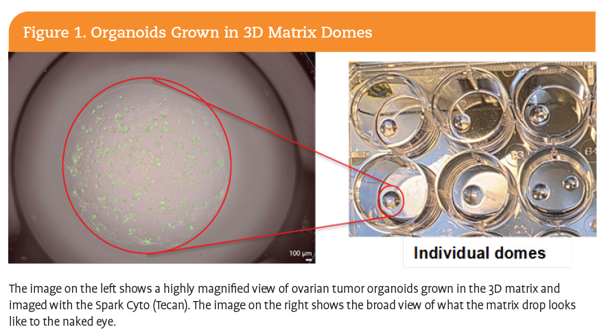
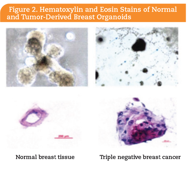
Organoids are formed by enzymatic and mechanical digestion of a patient’s tumor tissue into single cells that are mixed with a gel-like matrix to form a 3D dome structure. An example of organoids grown in 3D matrix domes is shown in Figure 1. A key step necessary for standardization of organoid studies is identifying appropriate growth media that support the expansion and maintenance of tumor cells outside the original microenvironment, while preserving the original characteristics of the tumor. This process allows the single cells to expand and grow into organoids that exhibit the genetically heterogeneous cells present in the initial tumor5 and maintain the molecular and histological characteristics of the original tissues,6 as seen in Figure 2. Often, just a small amount of tissue from a patient can yield multiple organoids that can be expanded for use in numerous studies.


Advantages of Organoids for Preclinical Drug Screening
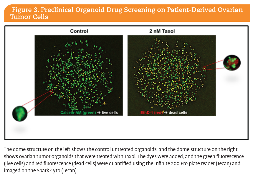
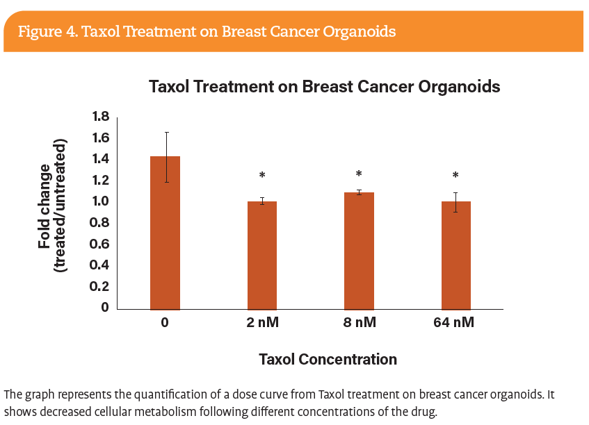
Patient-derived organoids provide a physiologically relevant plat form for evaluating drug response in diverse tumor types. Organoids are used to better understand the microenvironment cues and drug responses to preclinical and clinically available drugs. Importantly, drug responses can be evaluated in a relatively short time frame (2 to 3 weeks) compared to traditional preclinical models. For example, Figure 3 represents a drug treatment study that was performed on patient-derived ovarian organoids treated with or without Taxol for 5 days in a patient with ovarian cancer. Cellular viability was measured and imaged to quantify live and dead cells, respectively. Even with this short treatment time, tumor response is evident (Figure 4).


Organoid models can inform clinical trial design. As they maintain the genetic and phenotypic diversity observed in patient populations, organoid platforms can be used to identify characteristics and biomarkers of drug response to define patient selection. Furthermore, as most clinical trials occur in patients for whom front-line therapy has failed, organoid models provide the ability to screen drugs in treatment-naive tumors alongside early clinical trials in resistant patients.5 Additionally, the establishment of organoids from rare tumor types, in which suitable model systems were not previously available, can support efficacy assessments for rapid preclinical screening to guide therapeutic prioritization in smaller clinical trials.
Development of a Patient-Derived Organoid Core at an NCI-Funded Community Cancer Center
The majority of cancer patients in the US receive care at a community cancer center.7,8 The availability of a broad range of treatment-naive tumor types and the diverse patient population within the state of Delaware who receive care at the Helen F. Graham Cancer Center and Research Institute enabled the development of the Cawley Center organoid core and biobank. While organoid platforms have been established previously in academic research settings, this effort represents the first implementation of such an infrastructure at a community cancer center. Importantly, this setting captures real-world patient populations that reflect the demographic and clinical diversity observed in clinical trials and mirrors the rationale for conducting clinical trials in community cancer centers where routine care is delivered. This approach enhances the inevitable translational relevance of preclinical findings, improving clinical studies, lowering drug development costs, and impacting patient care.
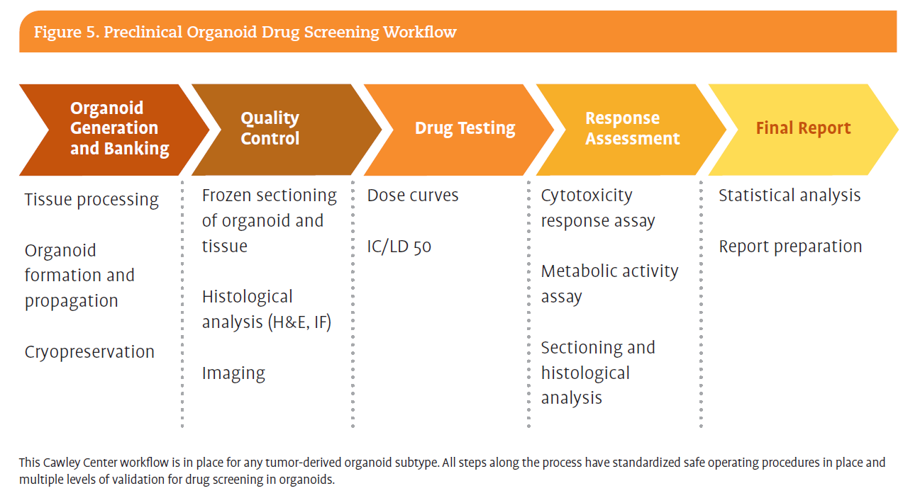
In this setting, the organoid core at the Cawley Center is focused on preclinical drug screening using patient-derived organoid models. As the facility continues to expand, this technology is essential for paving the way for new research avenues to target cancer more effectively. Figure 5 illustrates a robust organoid drug screening workflow developed by the organoid core team that is implemented on our internal research projects and with external collaborators, such as the 15-year Helen F. Graham Cancer Center and Research Institute partnership with the Ellen and Ronald Caplan Cancer Center at the Wistar Institute.9

Concluding Thoughts
The promise of precision medicine is on the horizon. Organoids are an evolving tool to increase accuracy and pace, and to reduce the cost of preclinical drug development. The ability to grow each patient’s tumor in 3D organoids allows us to fully capture the unique genetic changes that are lacking in current preclinical animal models. The organoid core at the Cawley Center, in collaboration with partners like the Wistar Institute, provides a clinically relevant platform to study new cancer biomarkers and treatments. This new platform will allow researchers and clinicians to better identify actionable drug targets and effectively model drug response, enabling cutting-edge research and shortening the time from bench to clinic. By combining advanced technology, strong community partnerships, and direct patient access, ChristianaCare and the Cawley Center are showing how translational cancer research can thrive in a community setting, making breakthroughs not only in the laboratory but also in patients lives.
Shirin R. Modarai, PhD, is a research scientist in Dr Sims-Mourtada’s lab who helped establish the organoid core. Lynn M. Opdenaker, PhD, is a research scientist and laboratory manager of the Cawley Center, and Jennifer Sims-Mourtada, PhD, is the associate director of the Cawley
Center and director of the Translational Breast Cancer Program at the Helen F. Graham Cancer Center. Nicholas J. Petrelli, MD, is the director of the Cawley Center and associate director of Translational Research at the Wistar Institute in Philadelphia, Pennsylvania.
Acknowledgements
We would like to acknowledge the Delaware Ovarian Cancer Foundation for their generous support in spearheading the ovarian cancer organoid research at
the Cawley Center and the Lisa Dean Moseley Foundation for their support of the Organoid Core.
References
1. Buntz B. NIH announces end to funding for animal-only studies. Accessed March 5, 2026. Drug Discovery & Development. Published July 7, 2025. Updated July 14, 2025. https://www.drugdiscoverytrends.com/nih-announces-end-to-funding-for-animal-only-studies/
2. US Food & Drug Administration. FDA announces plan to phase out animal testing requirement for monoclonal antibodies and other drugs. Published April 10, 2025. Accessed March 5, 2026. https://www.fda.gov/news-events/press-announcements/fda-announces-plan-phase-out-animal-testing-requirement-monoclonal-antibodies-and-other-drugs
3. Wouters OJ, McKee M, Luyten J. Estimated research and development investment needed to bring a new medicine to market, 2009-2018. JAMA. 2020;323(9):844-853. doi10.1001/jama2020.1166
4. Thorel L, Perr ard M, Florent R, et al. Patient-derived tumor organoids: a new avenue for preclinical research and precision medicine in oncol ogy. Exp Mol Med. 2024;56(7):1531-1551. doi.org/10.1038/s12276-024-01272-5
5. Nagle PW, Plukker JThM, Muijs CT, van Luijk P, Coppes RP. Patient-derived tumor organoids for prediction of cancer treatment response. Semin Cancer Biol. 2018;53:258-264. doi:10.1016/j.semcancer.2018.06.005
6. Dutta D, Heo I, Clevers H. Disease modeling in stem cell-derived 3D organoid systems. Trends Mol Med. 2017;23(5):393-410. doi:10.1016/j.molmed.2017.02.007
7. Petrelli NJ. A community cancer center program: getting to the next level. J Am Coll Surg. 2010;210(3):261-270. doi:10.1016/j.jamcollsurg.2009.11.015
8. Johnson MR, Clauser SB, O Brien DM, Beveridge JM, Kaluzny AD. Improving cancer care and expanding research in community hospitals: lessons from the National Cancer Institute Community Cancer Centers Program. Oncol Issues. 2011;26(1):26-28. https://www.academia.edu/118752134/Improving_Cancer_Care_and_amp_Expanding_Research_in_Community_Hospitals
9. Shao C, Indeglia A, Foster M, et al. Mutant p53 binds and controls estrogen receptor activity to drive endocrine resistance in ovarian cancer. Genes Dev. 2026;40(3-4):199-214. doi:10.1101/gad.352953.125




Recognizing the critical role advanced practice providers and allied health professionals play in the care continuum, the American Oncology Network provides infrastructure, leadership, and professional growth opportunities to empower these members of the cancer care team to practice at the top of their license.